
The Heart at a Glance
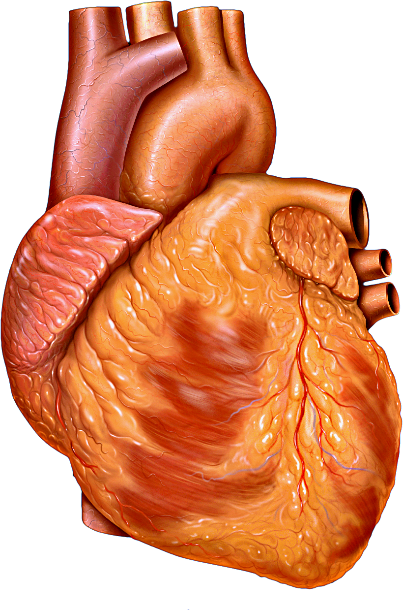
The human heart is a hollow, four-chambered muscular pump about the size of your fist, located in the mediastinum between the lungs and slightly left of center. It beats approximately 100,000 times per day — about 2.5 billion times over an average lifetime — and pumps roughly 2,000 gallons of blood daily. It never rests, never pauses, and begins beating just 22 days after conception.
The Four Chambers
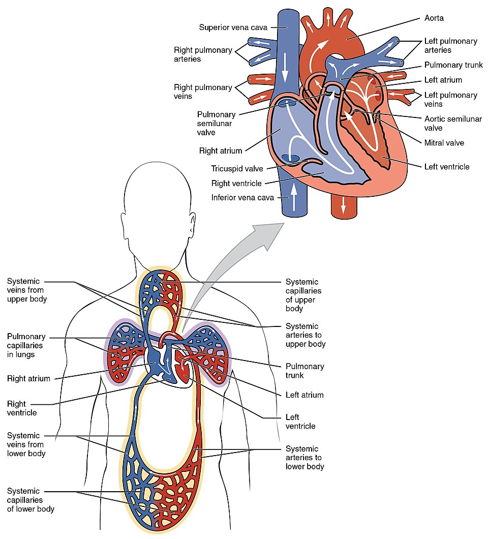
The heart is divided into a left side and a right side by the interventricular septum, and into upper and lower halves by the atrioventricular valves. This creates four chambers:
- Right Atrium: Receives deoxygenated blood returning from the body via the superior and inferior vena cavae. Also receives blood from the coronary sinus (coronary circulation return). Contains the SA node (pacemaker).
- Right Ventricle: Receives blood from the right atrium and pumps it through the pulmonary valve into the pulmonary artery, sending it to the lungs for oxygenation. Has a thinner wall than the left ventricle — it only pumps against pulmonary resistance (~25 mmHg).
- Left Atrium: Receives oxygenated blood returning from the lungs via the four pulmonary veins. Passes blood through the mitral valve to the left ventricle.
- Left Ventricle: The heart's workhorse. Pumps oxygenated blood through the aortic valve into the aorta and throughout the systemic circulation. Must generate pressures of 120+ mmHg. Its wall is 3× thicker than the right ventricle. The interventricular septum bulges toward the right ventricle.
The Four Valves: Ensuring One-Way Flow
Heart valves open and close with each heartbeat to ensure blood flows in one direction only — backward flow would be catastrophically inefficient.
Atrioventricular (AV) Valves — between atria and ventricles:
- Tricuspid Valve (Right AV): Has three cusps (leaflets). Prevents backflow from right ventricle to right atrium during systole. Supported by chordae tendineae attached to papillary muscles.
- Mitral (Bicuspid) Valve (Left AV): Has two cusps. Prevents backflow from left ventricle to left atrium. The most commonly diseased valve. Also supported by chordae tendineae.
Semilunar Valves — between ventricles and great vessels:
- Pulmonary Valve: Has three half-moon shaped cusps. Opens when right ventricular pressure exceeds pulmonary artery pressure. Prevents backflow from pulmonary artery to right ventricle during diastole.
- Aortic Valve: Has three cusps. Opens when left ventricular pressure exceeds aortic pressure. Closes during diastole, preventing aortic blood from re-entering the left ventricle. The coronary arteries originate just above this valve.
The "lub-dub" (S1–S2) sounds you hear through a stethoscope are the sounds of these valves snapping shut — not the muscle contracting.
The Cardiac Cycle
One complete heartbeat is the cardiac cycle, lasting approximately 0.8 seconds at rest (75 bpm):
- Atrial Systole (0.1 sec): Both atria contract simultaneously, pushing remaining blood into the ventricles (the "atrial kick" contributes ~15–25% of ventricular filling).
- Ventricular Systole (0.3 sec): Both ventricles contract. Pressure rises rapidly, closes the AV valves (S1 "lub"), then exceeds pulmonary and aortic pressures, opening the semilunar valves. Blood is ejected into the pulmonary artery and aorta.
- Ventricular Diastole (0.4 sec): Ventricles relax, pressure drops. Semilunar valves snap shut (S2 "dub"). AV valves open. Ventricles passively fill with blood from atria (about 70–80% of filling is passive, before the atrial kick).
Cardiac Output (CO) = Heart Rate (HR) × Stroke Volume (SV)
Normal resting values: 70 bpm × 70 mL = ~5 L/min. During maximal exercise in a trained athlete: up to 200 bpm × 120 mL = ~24 L/min. Ejection fraction (EF) — the percentage of end-diastolic volume pumped out — is normally 55–70%. EF below 40% indicates heart failure.
The Electrical Conduction System
The heartbeat is initiated by the heart itself — it does not require the brain or spinal cord. This property is called automaticity.
- SA Node (Sinoatrial Node): Located in the wall of the right atrium near the opening of the superior vena cava. The natural pacemaker, firing spontaneously at 60–100 times/minute in a healthy adult. Generates the action potential that starts each beat. The P wave on an EKG represents atrial depolarization spreading from the SA node.
- AV Node (Atrioventricular Node): Located at the junction of the atria and ventricles in the interatrial septum. The impulse slows here for approximately 0.1 seconds — this delay ensures the atria finish contracting and emptying into the ventricles before ventricular contraction begins.
- Bundle of His (AV Bundle): The only electrical connection between the atria and ventricles (the AV node provides the only electrical pathway through the otherwise electrically insulating fibrous skeleton). Conducts the impulse to the interventricular septum.
- Right and Left Bundle Branches: Travel down either side of the interventricular septum toward the apex of the heart.
- Purkinje Fibers: Spread throughout the ventricular walls, rapidly conducting the impulse to ventricular myocytes. This ensures the ventricles contract from the apex upward — efficiently squeezing blood toward the outflow tracts. The QRS complex on an EKG represents ventricular depolarization.
Blood Supply to the Heart: Coronary Circulation
The heart muscle is supplied by two coronary arteries that branch off the aorta just above the aortic valve:
- Left Coronary Artery (LCA): Divides into the left anterior descending (LAD) artery — which supplies the anterior left ventricle and interventricular septum — and the circumflex artery, which supplies the left atrium and posterior/lateral left ventricle.
- Right Coronary Artery (RCA): Supplies the right atrium, right ventricle, and typically the inferior left ventricle and the SA and AV nodes.
Blockage of the LAD artery is sometimes called the "widow maker" — it supplies the largest territory of myocardium. Coronary artery disease (atherosclerosis of these vessels) is the leading cause of death in the United States.
Clinical Connections
- Myocardial Infarction (Heart Attack): Prolonged ischemia (lack of blood flow) causes cardiomyocyte death. The affected area loses contractility, potentially causing arrhythmias and heart failure.
- Heart Failure: When the heart cannot pump enough blood to meet the body's needs. Left-sided failure causes pulmonary edema (fluid in lungs); right-sided failure causes peripheral edema (fluid in legs/ankles).
- Valve Disease: Stenosis (narrowed, stiff valve) creates turbulent flow (heart murmur) and increased workload. Regurgitation (leaky valve) allows backward flow, reducing efficiency.
- Arrhythmias: Abnormal electrical activity. Atrial fibrillation (irregular rapid atrial firing) is the most common. Ventricular fibrillation is immediately life-threatening.
Discussion (0)
Leave a Comment