
The Neuron: Structure Built for Communication
Neurons are the most electrically active cells in the body. A single human brain contains approximately 86 billion neurons, each capable of forming up to 10,000 synaptic connections — creating a network of roughly 100 trillion synapses. Everything you think, feel, perceive, and do is the result of electrochemical signals traveling through this network at speeds of up to 120 meters per second.
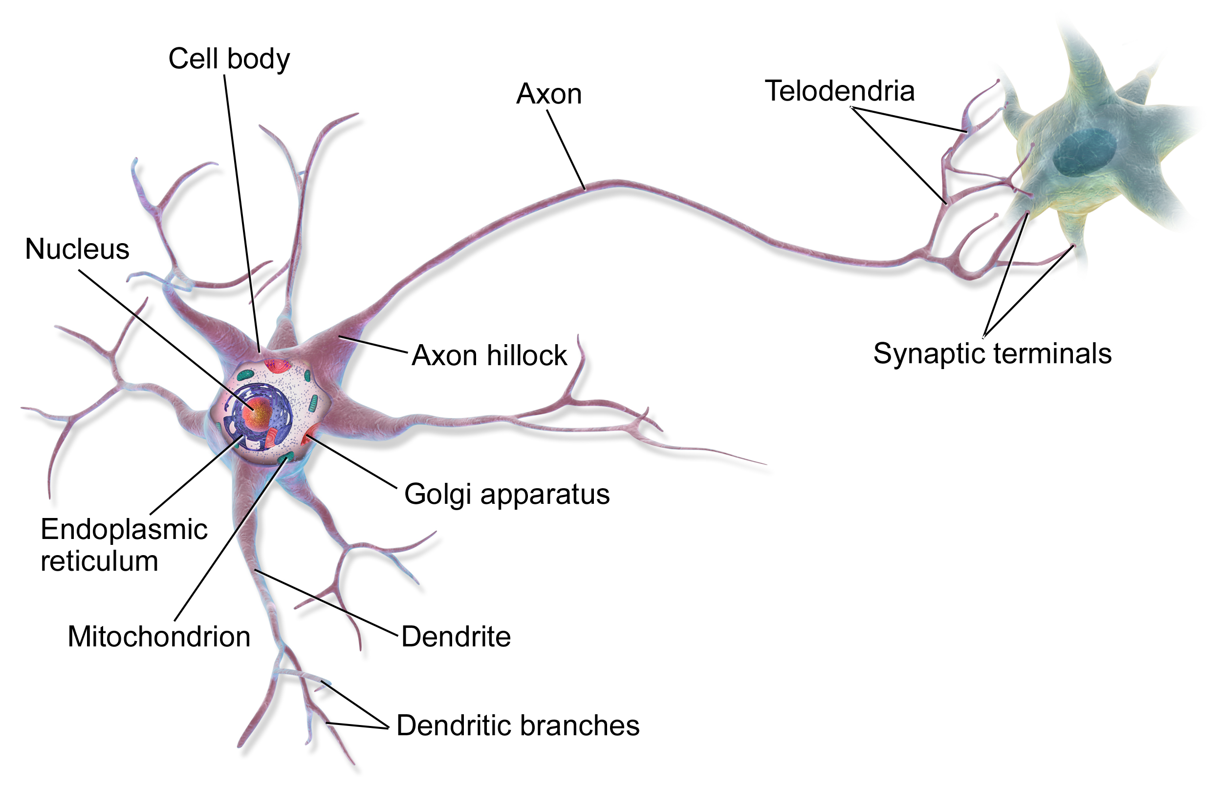
A typical neuron has four structurally distinct regions:
- Dendrites: Highly branched processes that extend from the cell body. They are the primary site for receiving incoming signals from other neurons. The more dendrites (and dendritic spines), the more inputs a neuron can integrate. Dendritic geometry can be modified by learning and experience — a process called structural plasticity.
- Cell Body (Soma): Contains the nucleus and most of the cell's organelles. Integrates all incoming signals from dendrites and determines whether to fire an action potential. Contains Nissl bodies (rough ER), which synthesize neurotransmitter-related proteins at a high rate.
- Axon: A single, long process that transmits signals away from the cell body toward other neurons or effector cells. Axons range from less than 1mm (local interneurons) to over 1 meter long (motor neurons from the spinal cord to the foot). The axon hillock — where the axon meets the cell body — is the site where action potentials are initiated (it has the highest density of voltage-gated Na⁺ channels).
- Axon Terminals (Synaptic Boutons): Enlarged endings of the axon that contain synaptic vesicles packed with neurotransmitters. When an action potential arrives, these terminals release neurotransmitters into the synapse.
Many axons are wrapped in a myelin sheath — multiple layers of lipid-rich membrane produced by Schwann cells (in the PNS) or oligodendrocytes (in the CNS). Myelin acts as an electrical insulator, dramatically increasing conduction velocity. Gaps in the myelin sheath occur at regular intervals called nodes of Ranvier. In demyelinating diseases like multiple sclerosis, the loss of myelin slows or blocks neural transmission, causing the diverse neurological symptoms of the disease.
Ion Gradients and the Resting Membrane Potential
All electrical signaling in neurons depends on concentration gradients of ions across the plasma membrane. At rest, the cytoplasm of a neuron is approximately –70 mV relative to the extracellular fluid. This resting membrane potential is maintained by two mechanisms working together:
1. The Na⁺/K⁺-ATPase Pump (Active Transport): This membrane protein uses one ATP molecule to move 3 Na⁺ ions out of the cell and 2 K⁺ ions in — against their concentration gradients. This creates and maintains the concentration gradients: high Na⁺ outside, high K⁺ inside. Because more positive charge is pumped out than in, the pump itself is electrogenic — it contributes slightly to the negative resting potential.
2. Selective Membrane Permeability (Leak Channels): At rest, the membrane is ~40× more permeable to K⁺ than Na⁺, due to open K⁺ leak channels. K⁺ diffuses down its concentration gradient (out of the cell), carrying positive charge out and leaving the interior relatively negative. Na⁺ wants to enter but cannot easily — its channels are mostly closed at rest. This unequal charge separation across the membrane is the resting membrane potential.
The key ion gradients at rest (intracellular vs. extracellular):
- Na⁺: 12 mM inside vs. 145 mM outside — strong inward driving force
- K⁺: 155 mM inside vs. 4 mM outside — strong outward driving force
- Cl⁻: 4–30 mM inside vs. 120 mM outside
- Large organic anions (proteins, phosphates): ~100 mM inside — impermeant, contribute to negative interior
The Action Potential: The Neural Signal
An action potential is a brief, rapid, stereotyped reversal of membrane potential that propagates down the axon without decrement. It is all-or-nothing: either the threshold is reached and a full action potential fires, or it does not. The magnitude of the signal does not vary (unlike a graded potential), but the frequency of action potentials encodes signal intensity.
Phases of the Action Potential:
- Stimulus and Graded Depolarization: A subthreshold depolarization (e.g., from an EPSP arriving at dendrites) spreads passively to the axon hillock. If the membrane potential reaches threshold (approximately –55 mV), voltage-gated Na⁺ channels open.
- Depolarization (Rising Phase): Voltage-gated Na⁺ channels open rapidly. Na⁺ rushes into the cell down its electrochemical gradient. The membrane potential rapidly rises from –70 mV to approximately +30 mV (overshoot). This is a positive feedback process — depolarization opens more channels, which causes more depolarization.
- Repolarization (Falling Phase): After approximately 0.5 ms at peak depolarization, Na⁺ channels inactivate (inactivation gate closes — they cannot reopen immediately). Simultaneously, voltage-gated K⁺ channels open (their activation is slower than Na⁺ channels). K⁺ flows rapidly out of the cell, restoring negative charge.
- Hyperpolarization (After-Hyperpolarization): K⁺ channels close slowly — K⁺ continues briefly to exit even as the membrane approaches –70 mV. The membrane transiently overshoots to approximately –80 mV. During this period, the neuron is in the relative refractory period — a stronger-than-normal stimulus can fire another action potential. During repolarization (while Na⁺ channels are inactivated), the neuron is in the absolute refractory period — no stimulus can fire another action potential regardless of intensity.
- Return to Resting Potential: K⁺ channels close, the Na⁺/K⁺ pump works to restore the ionic gradients, and the membrane returns to –70 mV.
Propagation: The action potential is self-propagating. Depolarization at one segment of the axon triggers depolarization in the adjacent segment (because current flows locally from the depolarized to the adjacent resting membrane, bringing it to threshold). It moves in one direction only (away from the cell body) because the previous segment is in its refractory period.
Saltatory Conduction: In myelinated axons, the action potential "jumps" from one node of Ranvier to the next, because the myelin prevents ion flow except at the nodes. This saltatory conduction (from Latin saltare, to jump) is dramatically faster than continuous conduction in unmyelinated axons and much more energy-efficient. Conduction velocity: unmyelinated C fibers ~0.5–2 m/s; myelinated A-alpha fibers ~70–120 m/s.
Synaptic Transmission: Neuron to Neuron
Communication between neurons occurs at synapses — specialized junctions between the axon terminal of a presynaptic neuron and the dendrite, cell body, or axon of a postsynaptic neuron. The vast majority of synapses in the human nervous system are chemical synapses — they use chemical messengers (neurotransmitters) to bridge the 20–40 nm synaptic cleft.
Sequence of chemical synaptic transmission:
- Action potential arrives at the axon terminal.
- Depolarization opens voltage-gated Ca²⁺ channels in the terminal membrane.
- Ca²⁺ influx triggers fusion of synaptic vesicles with the presynaptic membrane — exocytosis of neurotransmitters into the synaptic cleft. (Ca²⁺ is the critical link between electrical signal and chemical release.)
- Neurotransmitters diffuse across the synaptic cleft (20–40 nm — extremely fast, ~0.05 ms).
- Neurotransmitters bind to specific receptors on the postsynaptic membrane. These are ligand-gated ion channels (ionotropic) or G-protein coupled receptors (metabotropic).
- Ion channels open or close → change in postsynaptic membrane potential. Excitatory: Na⁺ influx → depolarization (EPSP). Inhibitory: Cl⁻ influx or K⁺ efflux → hyperpolarization (IPSP).
- Neurotransmitter is terminated by: enzymatic degradation (acetylcholinesterase cleaves ACh), reuptake into the presynaptic terminal (monoamine transporters), or diffusion away from the cleft.
Major Neurotransmitters:
- Acetylcholine (ACh): Motor neurons → skeletal muscle (NMJ); parasympathetic system; memory (Alzheimer's involves loss of cholinergic neurons).
- Dopamine: Reward, motivation, voluntary movement (Parkinson's: dopaminergic neuron loss); psychosis (excess dopamine in certain pathways).
- Serotonin: Mood, sleep, appetite, social behavior. Most antidepressants (SSRIs) block serotonin reuptake.
- GABA (γ-aminobutyric acid): The main inhibitory neurotransmitter in the brain. Benzodiazepines (anti-anxiety drugs) and alcohol enhance GABA activity. Reduced GABA activity is associated with anxiety and seizures.
- Glutamate: The main excitatory neurotransmitter. Essential for learning and memory (LTP at NMDA receptors). Excess glutamate (excitotoxicity) damages neurons after stroke or trauma.
- Norepinephrine: Arousal, attention, fight-or-flight; sympathetic postganglionic neurons. Many antidepressants also enhance norepinephrine.

Discussion (0)
Leave a Comment